Cell Proliferation and Viability Assays
Cell-Based Assays

Cell Proliferation and Cell Viability Assay Services

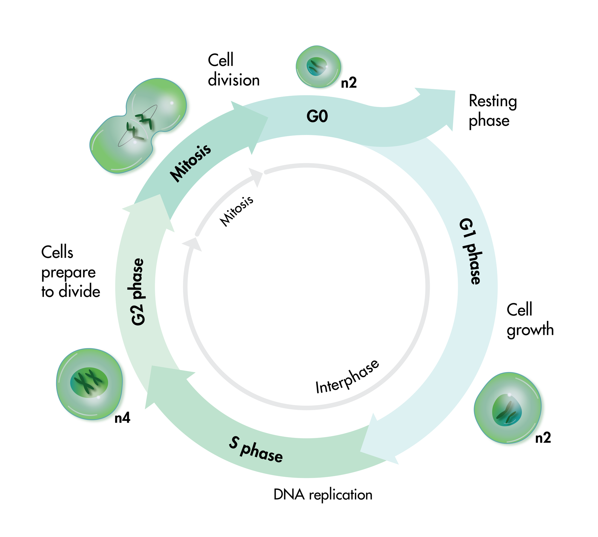
Cell viability and cell proliferation assays are indispensable for any cancer drug discovery project. Cell proliferation assays are widely used as an initial test for the therapeutic efficacy of anti-cancer agents.

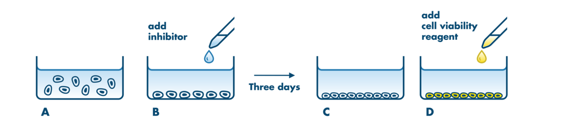
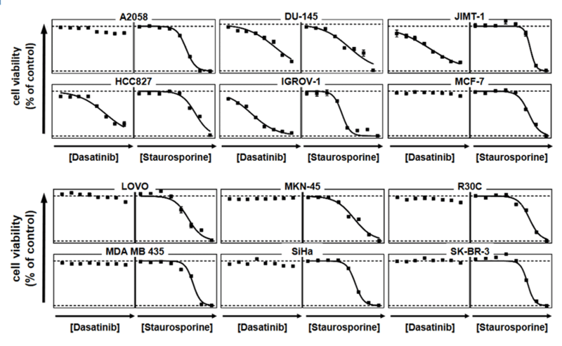
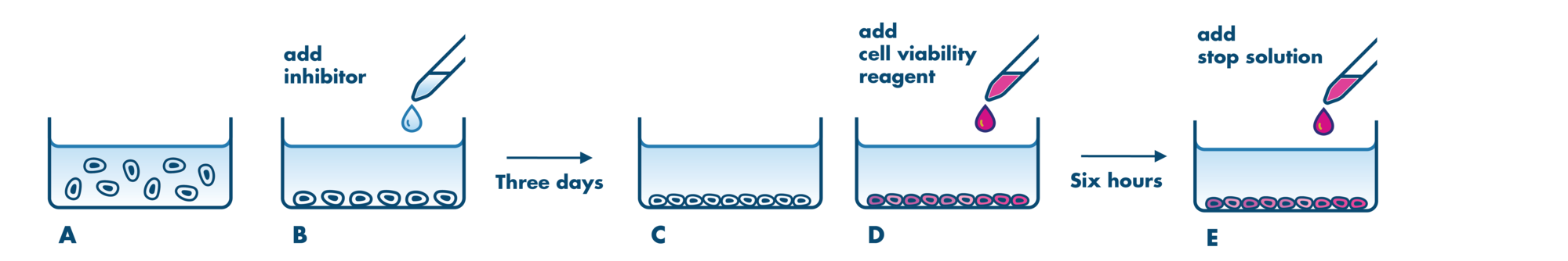
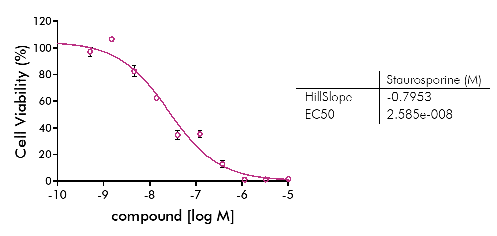
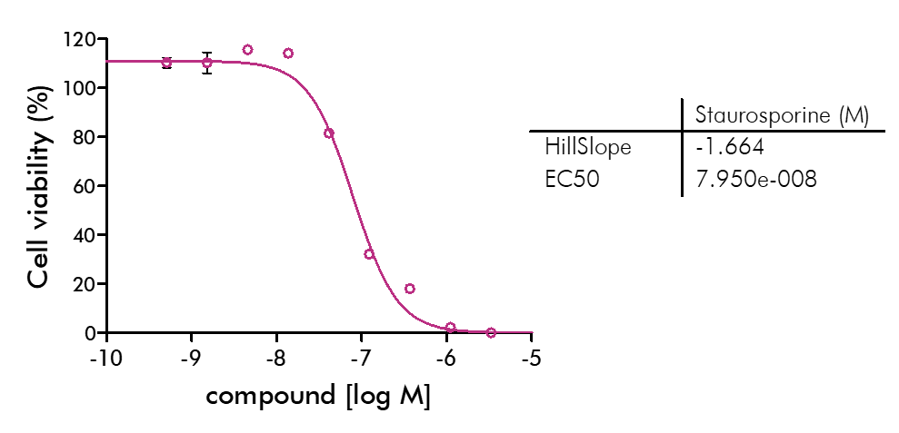
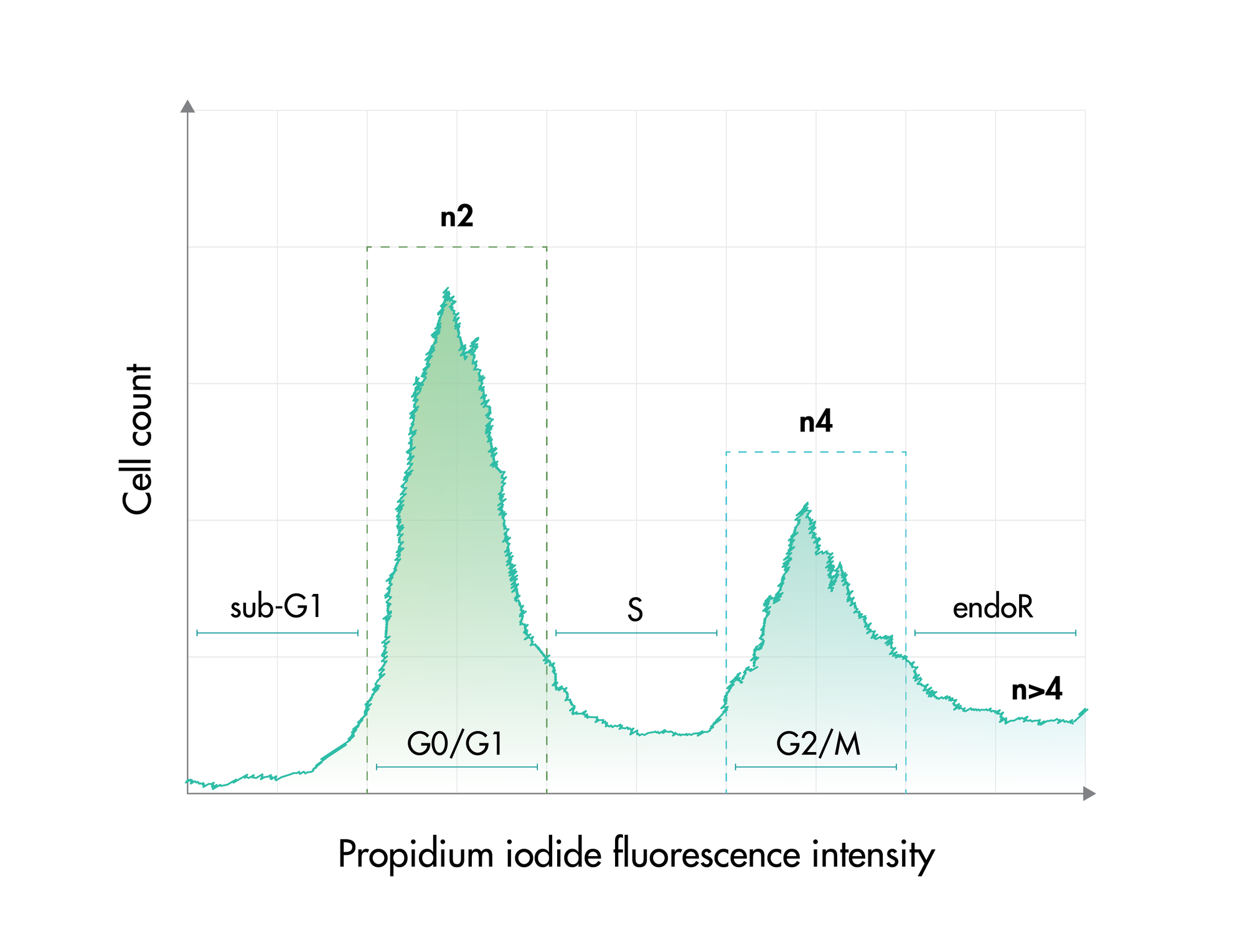
For analysis of the impact of anti-cancer drugs, the most common cellular phenotypic assay is the 2D cell proliferation assay in which tumor cells are growing in suspension culture or as monolayers in cell culture plates. Our standard assay format Cell Titer-Glo which allows the testing of large numbers of compounds in a fast and cost-efficient fashion. Due to its simplistic setup, the cell proliferation assay is generally regarded as a pretest to more sophisticated assays such as apoptosis or 3D soft agar assays, which are also available at Reaction Biology.

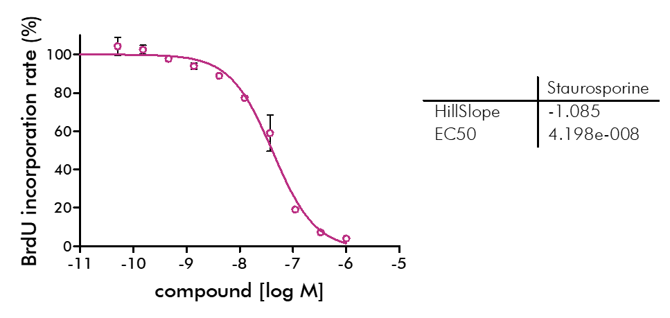
  • Beside Cell Titer-Glo, we offer potency testing of new drugs on cell proliferation and viability with a variety of alternative assay formats are available including ATPlite, CyQUANT, BrdU incorporation, MTT, Alamar Blue, and WST-8 readouts as well as real-time kinetics measured via IncuCyte.
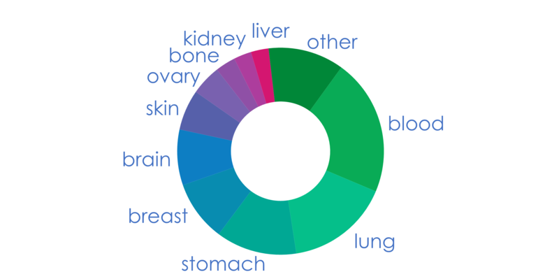
  • Regular large tumor cell panel screening is available with our ProLiFiler™ screening service.
  • Our 2D cell proliferation assays allow for drug incubation times up to 3 days. For projects that may require incubation times >3 days to develop a drug response, we recommend our 3D Tumor Spheroid Assay Service or the Soft Agar Assay Service which are well suited to this application. Please contact us to discuss the details.

Our gold standard assay formats, the large cell line panel, and exceptional service make us the choice contract research organization for quality cell proliferation assays. Contact our global business development team today to discuss your contract research needs.

253 tumor cell lines

253 human tumor cell lines are available for drug testing in our cell proliferation and viability assays.

Cell lines that are not in our panel can be established according to the specific research needs of our customers.

Please inquire for a list of our cell lines.

Cell Lines Graph

Cell Lines Graph

Cell Proliferation Assay Details

  • CellTiter-Glo
  • MTT assay
  • CyQUANT assay
  • BrdU Incorporation Assay
  • IncuCyte
  • Cell Cycle Analysis
  • Drug Combination Testing
  • Assay Details
CellTiter-Glo

CellTiter-Glo

Goal: Testing of the cytotoxic or anti-proliferative capacity of compounds based on the metabolic activity of live cells.

Assay principle: Promega‘s CellTiter-Glo dye is used to determine the number of cells in cell culture by generation of a luminescent signal proportional to the amount of ATP present based on the light emitted by luciferase during ATP-dependent oxidation of luciferin.

Assay procedure:

assay principle of the cellular proliferation assay performed with celltiter-glo

Cells are grown in multi-well plates (A) and incubated with the compound for 72 hours (B, C). The number of viable cells is determined by luminescence readout after the addition of CellTiter-Glo dye (D).

Details:

•This is the go-to assay for first-in-cell data for new test compounds.

•The assay is performed with a homogenous add-mix-measure format which does not require wash steps allowing for accurate results.

•The dye is stable for several hours which makes the assay less susceptible to human readout errors.

•The procedure is performed in 96 or 384 well format and is, therefore, high-throughput compatible.

•Available for ProLiFiler™ – Cell Line Panel Screening assay on a set of 160 tumor cell lines

Available alternative: ATPlite (Perkin Elmer)

Example data CellTiter-Glo assay:

cell proliferation assay example data

A tyrosine kinase inhibitor and staurosporine were tested for proliferation inhibition of 12 cancer cell lines. 72 h after compound addition, live cells were quantified via CellTiter-Glo. IC50 values are expressed as a percentage of proliferation in the presence of solvent alone (100% = high control) compared with cells treated with 1E-5M staurosporine (0% = low control).

MTT assay

MTT Assay

Goal: Testing of the cytotoxic or anti-proliferative capacity of compounds based on the metabolic activity of live cells.

Assay principle: The MTT assay is useful to determine the number of cells in cell culture via an NADH-dependent reduction of yellow tetrazolium salt (3-(4,5-Dimethylthiazol-2-yl)-2,5-diphenyltetrazolium bromide) to purple formazan crystals via colorimetric readout.

Assay procedure:

MTT Assay

Cells are grown in multi-well plates (A) and incubated with compound for 48 hours (B, C). MTT dye solution is added (D). After a 6 hour incubation period a solubilization/stop solution is added (E) and the number of viable cells is determined based on the colorimetric readout by Envision 2104 Multilabel Reader.

Available alternative assays:

MTS and WST-8 assays are advanced versions of the MTT assay. The WST-8 assay is performed with WST-8 or CCK-8 solution (Dojindo) which is not cytotoxic and allows additional readouts. The MTS assay is based on the reduction of MTS tetrazolium by viable mammalian cells to generate a colored formazan dye that is soluble in cell culture media.

Alamar Blue assays are based on the cell-permeable and non-toxic resazurin which is a blue dye that undergoes a color change in response to a metabolic reduction in live cells. The resulting resorufin is pink and can be detected via fluorescence or colorimetric readout.

Example data MTT assay:

cMTT assay example

2000 KMS-11 human multiple myeloma cells were seeded into each well of a 384-well plate. Cells were treated with the indicated concentrations of reference compound staurosporine for 48 hours. MTT Dye Solution was added to each well and incubated for 6 hours at 37ºC until formazan was produced in the cells. Solubilization/Stop Solution was added to each well to dissolve the formazan crystals. The absorbance of each sample was measured using Envision 2104 Multilabel Reader. The IC50 value was calculated and IC50 curve was plotted using the GraphPad Prism program based on a sigmoidal dose-response equation.

Example data WST-8 assay:

WST-8 assay example

5000 HepG2 human hepatocellular cancer cells were seeded into each well of a 384-well plate. Cells were treated with the indicated concentrations of reference compound staurosporine for 72 hours. CCK-8 Solution (product from Dojindo) was added to each well and incubated at 37°C for 4 hours. Absorbance of each sample was measured using Envision 2104 Multilabel Reader. The IC50 value was calculated and IC50 curve was plotted using the GraphPad Prism program based on a sigmoidal dose-response equation.

CyQUANT assay

CyQUANT Assay

Goal: Determination viable cell numbers by determination of DNA content.

Assay principle: The CyQUANT Direct Cell Proliferation Assay is based on a DNA-binding dye which is cell permeable. Dead cells are blocked from staining by a masking dye.

Assay procedure:

Cell Proliferation assay

Cells are grown in multi-well plates (A) and incubated with compound for 48 hours (B, C). CyQUANT Detection Reagent is added to the cells and fluorescence is quantified.

Details:

•This assay provides a readout that is independent of metabolic state and cell cycle.

•It is possible to multiplex with spectrally distinct fluorescent probes or luminescence assay kits because cells are not lysed.

Example data CyQUANT assay:

CyQUANT assay example

2000 Jurkat human T cell leukemia cells were seeded into each well of a 384-well plate. Cells were treated with the indicated concentrations of reference compound staurosporine for 48 hours. CyQUANT Assay Detection Reagent was added to each well and incubated for 60 minutes at 37ºC. Fluorescence was measured by BioTek Synergy4 Microplate Reader. The IC50 value was calculated and IC50 curve was plotted using the GraphPad Prism 4 program based on a sigmoidal dose-response equation.

BrdU Incorporation Assay

BrdU Incorporation Assay

Goal: Testing of the anti-proliferative effects of compounds by quantification of replicating cells.

Assay principle: BrdU (Bromodeoxyuridine) is a pyrimidine analog that incorporates into cellular DNA during DNA synthesis instead of thymidine. BrdU can than be detected via a BrdU-specific antibody.

Assay procedure:

BrdU Assay

Cells are grown in multi-well plates (A) and incubated with compound for 72 hours (B, C). BrdU is added to the cells (D). After 24 hours, fixing/denaturing solution is added (E) and BrdU is detected via ELISA (F).

Details:

The BrdU Incorporation Assay gives insight into the action of compounds distinguishing the anti-proliferative effects vs. cytotoxic effects.

Example data for BrdU assay:

BrdU assay example

BT-474 breast cancer cells were seeded into each well of a 96-well plate. The cells were treated with the indicated concentrations of reference compound staurosporine for 72 hours. The BrdU incorporation into cellular DNA during cell proliferation was measured using BrdU ELISA assay. Absorbance signal was recorded by Envision 2104 Multilabel Reader. The IC50 value was calculated and IC50 curve was plotted using the GraphPad Prism program based on a sigmoidal dose-response equation.

IncuCyte

IncuCyte

Goal: Determination of the anti-proliferative or cytotoxic effects of compounds on cell culture with a label-free protocol and the option to acquire kinetic data.

Assay principle: The IncuCyte S3 Live-Cell Analysis system measures the amount of cells over a period of time via time-lapse imaging. Options for measurements are cell confluency and label free cell counting as well as counting of nuclei or fluorescently labelled cells.

Assay procedure:

IncuCyte assay

Cells are grown in multi-well plates (A) and incubated with compound for a variable time (B). IncuCyte will take images of the cell culture for live monitoring cell confluence or count cells (C).

Details:

•Assessment of the kinetics of the growth inhibition or cytotoxic effects of a compound

•Option for multiplexing with apoptosis or cytotoxicity reagents

•Co-culture studies with fluorescently labelled cells are possible

•Suitable for low and medium throughput

Cell Cycle Analysis

Cell Cycle Analysis

Goal: Cell cycle analysis is helpful in distinguishing cytotoxic and anti-proliferative effects of compounds when treating tumor cells. Also, the disruption of the cell cycle is one of the key strategies when developing new cancer drugs making the examination of the cell cycle a standard procedure in cancer drug discovery.

Assay principle: DNA content quantification with propidium iodide enables the measurement of cell frequencies in the cell cycle phases.

Cell Cycle

Cell cycle graph

The DNA content changes during cell cycle:

  1. Apoptotic cell fragments in sub-G1
  2. Diploid cells during G0 and G1 phases
  3. The DNA content increases during the S phase
  4. Tetraploid status in G2 and mitotic cells
  5. EndoR comprises endoreduplicated cells that have multiple nuclei due to interrupted cytokinesis

Example results:

PI Intensity

Tumor cells were incubated with reference compound Nocodazole for 24 hours. Cells were harvested by trypsination, and cells and supernatant were fixed with 90% methanol. After rehydration and RNAse A treatment, cells were stained with propidium iodide (PI) and analyzed via flow cytometry (above). The results were displayed below as % change of cell population in the cell cycle phases as compared to vehicle control.

Nocodazole cycle graph

Nocodazole treatment resulted in a reduction of tumor cells in G0/G1 phase and a high number of cells in G2/M phase indicating cell cycle arrest before or during mitosis.

Drug Combination Testing

Drug Combination Testing

Combining drugs that affect synergistically-acting targets is a promising strategy for drug discovery.

Depending on the drugs’ effectiveness and their mechanisms of action, different approaches are being used to determine combinatorial drug effects.

At Reaction Biology, we can perform
(i) a one-sided approach, i.e., the concentration of one drug is set, and the concentration of the other drug changes or
(ii) a two-sided approach, i.e., both drugs are tested with several concentrations in a checkerboard matrix setup.

Combination testing is available for Cell Proliferation Assay, ProLiFilerTM, Soft Agar Assay, Migration Assay, and 3D Tumor Spheroid Assay.

Assay Details

Assay details for CellTiter-Glo readout.

Setup: IC50 value determination and single concentration testing. Free choice for screening of the cell lines of your choice or panel screening via ProLiFiler™.

Controls: DMSO only treatment serve as high control. Staurosporine treatment (at 1×10⁻⁵M) serves as low control. In every project, testing of a standard reference compound is included in IC50 format.

Custom assay development: Custom-tailored assays can be performed. Cell lines that are not part of the list, can be established according to the client’s research needs.

Report: A detailed report including assay conditions, methodology, and comprehensive evaluation of data as well as raw data for each analysis will be provided.

Turnaround time for standard studies is about 3 weeks. Expedited scheduling and data delivery can be arranged prior to study commencement.

Compound Information: For CellTiter-Glo assays please download our detailed sample requirements and shipping instructions for cell-based assays (German Facility) here. For compound requirements of additional assay formats, please inquire.

Screening facility: CellTiter-Glo assays are performed at our German site. Additional readout formats are performed at our US site.